You are here

Drafts

Introcution, results for method draft

Submitted by jkswanson on Sat, 10/06/2018 - 16:57

Introduction

    The goal of this project is to create a multi panel scientific figure that is about a spider web, then write the methods of the procedure followed to create it.  Someone else will then follow the original methods and attempt to replicate the original image.

 Results

   Right off the bat the biggest difference seen is the web itself and the tree and are of the photo are all completely different.  The tree of the replicate is about 100 feet from the original tree. In figure A the webs themselves are different in size and visibility with the original being much larger and much more visible.  The original contains a dark purple folder behind the web to help it be more visible which is lacking in the replicate. There is also a leaf and other debris that are in the web in the original but not in the replicate.  The original image also contains a thumb and shouldve had a measuring tape at the bottom, but the editing messed it up, the replicate contains neither. Moving to figure A the map, they are different in size, the original being more zoomed in than the replicate.  The area circled in the replica is smaller than the original and black font is used as opposed to red in the original. The letters used to distinguish each figure are large black text in the original while the replica used drawn black letters. The positioning of these letters in the corner of each figure is different between the two figures.  In figure C the pictures are just different trees and areas. The entire panel of the original creates a tall rectangle that has each figure outlined in white. The replicat is not outlined and creates an elongated rectangle as the images are just placed in a row as opposed to figure A as a tall rectangle and B and C two squares stacked on top of eachother.  


Methods (rewritten)

Submitted by fmillanaj on Sat, 10/06/2018 - 13:55

To find a spiderweb on the UMass campus, I had to go through many trials and errors. The first spiderweb I found was too small for my phone to recognize. I had to go search for a bigger (more-defined) spider web. After a few days of searching, I found a spider web on the side of the Lederle Graduate Research center. On the section facing the main road (N Pleasant St), there was a spider web at about hip height. Photographing this web was quite difficult. I had to try to photograph it at several angles, with and without flash. I found that flash worked the best in making the web visible on in my photo. I had to angle the phone so that the camera was parallel to the main part of the spider web. (Addendum --- In addition to the spider web picture, I found a picture of the Lederle Graduate Research Center building (from the UMass Amherst website -- https://www.umass.edu/llc/lcc/lcc) on which the spider-web was on. This was to better show the location of the spider web in addition to providing a guide as to which side of the building the web was on.

    To create the figure, I gathered the location of the spider web on openmaps.eu, my photos of the spider web, and the picture of the Lederle Graduate building and put them in the inkscape app. I put the map on the top, the photo of the web on the bottom rights side, and the Graduate Building on the left bottom side. Then, I created labels to point out where the location of the spider web was, on both the map, and the pictures of where the web was. I labeled the map A in red font, the picture on the bottom right B in red font, and the picture on the bottom left C in red font. The organization of this was mainly to highlight the locations, starting from the furthest, the map to the closes, an actual picture of the spider web.


Psephurus and Polydon

Submitted by mtracy on Sat, 10/06/2018 - 00:08

Psephurus, or chinese paddlefish, is a large cartilaginous fish of the order acipenserifomres. These live in freshwater rivers, such as the Yangtze and its associated lakes. Historically, Psephurus could reach 7 meters in length. Unfortunately over time, these large fish were fished nearly out of existence. Additionally it was a major victim of the construction of the three gorges dam, as the migration that takens place between mating seasons could no longer take place. The last known specimen was killed illegally in 2007 and was only 3.6m long. No other Psephurus have been identified since.

The North American cousin to Psephurus is known as Polydon. Polydon lives in large freshwater rives as well, such as the Misissippi and Ohio rivers. Its long snout is covered with sensitive electrosensors, which can be used to detect swarms of plankton. The fish will swim forcefully through the water, injesting the plankton which are filtered through the gill rakes. Plankton are diverted to the fish's stomach, while water exits through the gill slits. Unlike Psesphurus, Polydon does not get as large; only about 2 meters in length. Unfortunately, much like Psesphurus, Polydon is a victim of overfishing, and due to dams can only be sustained by captive breeding.

Observations and Inferences

Submitted by yurigarcia on Fri, 10/05/2018 - 15:38

In the original picture the spiderweb is in the middle, the environment is in the middle and the map is on the right. On the replicate the spider web is on the left, the environment is on the middle and the map is on the right place. The arrow is pointing at the spiderweb on the original and in the replicate is pointing at the environment. The labeling letters in the original are in the picture and in the replicate are underneath of the pictures.  The original pictures are more zoom in and the replicate is taken from a far-away angle. Although it was taken in the same location it doesn’t show the spiderweb picture as the original. The maps are different in the original the ISB is emphasized and zoom in and in the replicate the map doesn’t specify the location.

Methods Draft

Submitted by jnduggan on Fri, 10/05/2018 - 13:48

The pictures themselves also differ from original to replicate.  Due to the quality of the pictures and lack of arrows on the environment picture of the replica, it is difficult to tell if the two spider webs are the same. The appearance of the stones relative to the spider web is different between the original and replicate in the close up picture.  In the environmental picture of the original figure, the curb and a blue building are visible in the background, but in the replica picture an orange building is visible and the curb is not. There is also a different number of posts on the fence visible in the replica vs. the original.

The pictures in the replica figure of the environment and close up have water on the stones and a relatively dark complexion, while the pictures of the environment and the spider web have a bright complexion and no water on the stones.  


discussion draft

Submitted by msalvucci on Fri, 10/05/2018 - 13:03

Observations of the replicated figure panel indicate many differences in the photographs between the two figure panels. The fact that the replicated picture shows a different amount of the tree next to the stair case suggests that the picture was taken a different angle than the original image. It was also observed that the picture showed more of the background of the spiderweb. From this difference, it is inferred that the photograph was taken from farther away in the replicate photo, thus, showing part of the student’s hands and more of the background behind the spiderweb.

The quality of the photographs taken are different between the original and replicated figure panels. The factor that’s most likely creating these differences in the lighting of the photographs would be the time of day that these pictures were taken.The weather also could be a factor affecting the picture quality, as the original pictures were taken on an overcast, cloudy day. This is most likely points to the camera exposure and focus.

The observed differences between the original and replicated figure panels are likely due to the in discrepancies in the methods. For example, the font size and type were different because the methods failed to completely describe the formatting. It is also inferred that a different version of Microsoft Word was used to create the replicate figure, as this would explain why the formatting differed.


Key draft

Submitted by eehardy on Fri, 10/05/2018 - 12:29
Key
A) Location of the Student Union on the Umass Campus Map. Student Union is circled in red. B) Front of the student Union. There are four different plants in front of the Student Union. The one of choice is circled in red. C) Plant pot on which the spider was located. Red arrows indicate the specific spot on the pot where the spider is located. D) Close up image of the spider.

Methods draft

Submitted by angelasalaza on Fri, 10/05/2018 - 12:14

The experiment provided instruction as to how the panel format should be presented in Swan’s method section. The instructor's guide began with opening Inkscape and begin importing the pictures onto the browser. That became a difficulty as Inkscape did not run on the current model of the experiment's designated Macbook pro. To accommodate the procedure the photographs were uploaded in the computer laboratory of Morrill two via USB import. The photographs were directed from the instructor’s method to be specific when placing in units of measurement by Inkscape and organize photographs in catalog order from the different views of the spider web and location. From the instructor’s guide, the experiment lacked consistency with the direction of measurement insertion, third-party help was needed to advance through the panel creation. The photographs were uploaded separately from the computer saved files and imported as images on to Inkscape. Each photograph is identified to serve purpose in this experiment the photograph of the stairwell demonstrated point of view and location in reference to the spider web, the screenshot of Live Maps identified the exact coordinates of the observer’s location and the dollar bill was used as a scale to identify size of the spider web.

The instructions followed a set of specific placement and measurements for each individual picture.  The x-axis location of the stairwell photograph measured 0.394 units across the x-axis, 219.951units above the y-axis, width of the picture measured 340.454 units, and height measured 250 units. All units of measurements were placed into the coordinate bar located at the top of the browser. Similar procedure was followed with the remaining two photographs but with varying units of measure. The photograph containing the staircase and spider web measured  0.394 units across the x-axis, 0.0 units above the y-axis, 340.454 units for width of the photograph and 219.951 units for the height of the photograph the screenshotted map measured 0.394 units across the x-axis, 0.0 above the y-axis, 340.454 units for width, and 219.951 units for height. All photographs were labeled at the top left corner with a white box outlined in black. Each box contained different measurements as to its placement the stairwell photograph was labeled with box A located at 0 units x-axis, 428.781units y-axis, 42.0 units for width of the picture, and 41.563 units for height of the unit, the screenshot of the map was labeled B with the measurements of 0.0 units across the x-axis, 178.781 units above the y-axis, 42.9 units in width, and 41.563 units in height and box C measured 340.454 units across the x-axis, 428.781 units above the y-axis, 42.0  units in width and 41.563 units in height. The last figure to be inserted had been an arrow measured at 245.877 units across the x-axis, 265.821 above the y-axis, 45.519 units in width of the picture, and 46.193 units in height on the photograph containing the stairwell photo pointing toward the placement of the spider web.

No observation could be made regarding differences between the original and replicate photograph of the spider web as the instructor did not upload the original photograph into the research project. Though it can be inferred that the replicate was inaccurate as it is too small to be viewed by the reader. The experiment performed was flawed as the instructions for finding the indicated location of the spider web was unclear no markers were used to indicate the correct location or placement of the spider web. Instructor’s directions provided a vague basis as to what stairwell is the correct one. The directions should have required counting each staircase to know which is the exact one used.
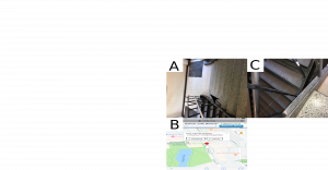
 

Figure 1 replicate


second part of methods intro

Submitted by eehardy on Fri, 10/05/2018 - 12:01

My picture was of a spider nestled between the rungs on a plant pot right in front of the Student Union on Campus. I selected this area because it was pretty close to the Biology Computer Resource Center, which I used as a reference point when describing the directions to the Student Union. I used the Biology Computer Resource Center as a reference point because that is where our class takes place, so I knew that the student following my Method would know its location, thus allowing them to replicate my procedure with ease. One factor that I knew I had to control was the location of the pot where the spider was. There were 4 different pots in front of the Student Union, so I had to specify which one by stating that it was the one on the end nearest the Lincoln Campus Center. I also needed to control where the picture was taken on the pot, since it was a large pot with several potential places that a spider could nestle. So I specified that the spider web was located between the vertical ridges on the pot, right above a patch of vegetation that was growing on the ground beneath the pot. Another variable that I sought to control was the platform for the creation of my image. I knew that most of the class was going to be using Inkscape, but I could not download Inkscape. I looked into Microsoft Word, mainly because it is a very popular platform, so I figured that the person replicating my image would likely already have Word. If they were not able to use the same platform as me, they would not be able to follow my procedure as precisely. I also specified the different colors of the font that I used, and when I put letters in bold. In general, the more detailed I was in my Methods, the more I controlled the different variables. 

MP- Abstract (Rough)

Submitted by cwcasey on Fri, 10/05/2018 - 11:47

Science is about imperfections and this project highlights that montra to a tee. Blindly assembling a scientific figure off of someone else's methods is a daunting task and is sure to generate interesting results  similar to those put forth later in this paper. After receiving the replicate figure, it was evident that the differences between the two figures were due to human error or natural differences. Once I was finished going through and seeing why and how these differences arose, it was important to comprise a short briefing that illustrated the explanations as to how the methods could have been vague and misunderstood. The remainder of this paper will serve as a guideline for how important attention to detail based on the differences between Figure one and Figure Two.


Pages

Subscribe to RSS - Drafts